The anatomy and biomechanics of genital prolapse.
Pelvic floor mri radiographics.
Imaging in the perioperative setting can be used as an objective measure after pelvic floor intervention to document anatomic and functional changes 8 12.
However imaging findings may not always correlate well with the clinical findings and symptoms 13 14.
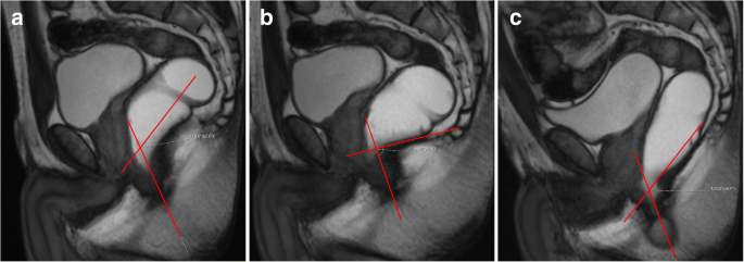
Dynamic analysis and evaluation of patients before and after surgical repair.
Mri of pelvic floor dysfunction.
Mr imaging based assessment of the female pelvic floor.
Paraurethral ligaments arrowheads in a which arise from the lateral wall of the urethra u.
28 fielding jr versi e mulkern rv lerner mh griffiths da jolesz fa.
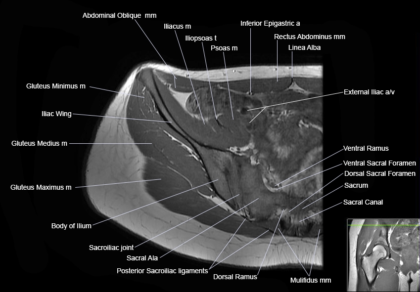
Normal female pelvic floor anatomy.
The periurethral ligaments arrows which arise from the pu borectalis muscle.
Practical mr imaging of female pelvic floor weakness.
Pelvic floor failure is a common disorder that affects 23 7 of women in the united states with a prevalence of 9 7 49 7 that increases with age one in nine women will undergo an invasive procedure for treatment of urinary incontinence or pelvic organ prolapse with 30 requiring additional surgery for symptom recurrence by 80 years of age.
Mr imaging of pelvic floor.
Axial t2 weighted mr images show the ligaments that support the female urethra at superior a and inferior b levels.
Colaiacomo mc masselli g polettini e et al.
Clin obstet gynecol 1993.
Top magn reson imaging.
Boyadzhyan l raman ss raz s.
Role of static and dynamic mr imaging in surgical pelvic floor dysfunction.
36 stoker j halligan s bartram c.
Role of static and dynamic mr imaging in surgical pelvic floor dysfunction.
Boyadzhyan l raman ss raz s.
Mri of pelvic floor dysfunction esur and esgar recommendations.
Magnetic resonance imaging of pelvic floor relaxation.
Dynamic mr imaging of the pelvic floor performed with patient sitting in an open magnet unit versus with patient supine in a.
The female pelvic floor is composed of the vulva levator ani muscle deep to it and the hollow viscera urethra vagina and rectum that penetrate through the levator ani at the midline 7 8 the supporting framework is the pelvic bony ring pubic rami ischium ilium sacrum and coccyx.
Imaging can play an additional role in the postoperative setting in the evaluation.
Mr imaging of the female pelvic floor in the supine and upright positions.
Dynamic mr imaging of the pelvic floor.
Ajr am j roentgenol.