Pelvic Floor Muscles Ct Scan
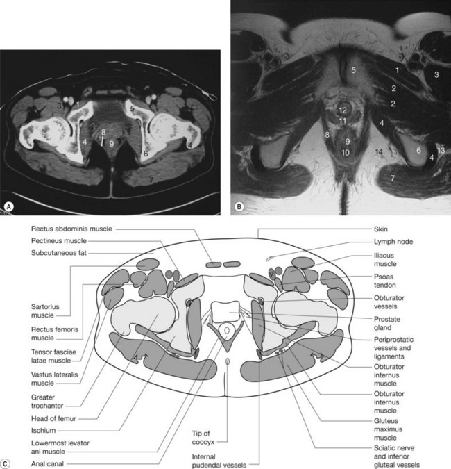
The Pelvis Radiology Key
Http Pdf Posterng Netkey At Download Index Php Congress Ecr2014 Module Get Pdf By Id Poster Id 119484

Above Shows A Number Of Possible Measurements Using Mri Imaging A Download Scientific Diagram
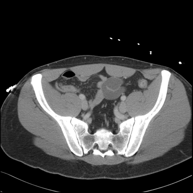
Ct Abdomen Pelvis Lower Axial Labeling Questions Radiology Case Radiopaedia Org

Figure 3 From Obturator Hernia Semantic Scholar
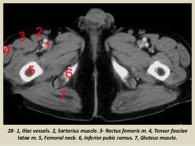
Presentation1 Pptx Ct Normal Anatomy Of The Abdomen And Pelvis
An abdominal ct takes pictures of your abdomen.
Pelvic floor muscles ct scan.

Predictive Role Of Measurement Of Pelvic Floor Muscle Thickness With Static Mri In Stress And Mixed Urinary Incontinence Semantic Scholar

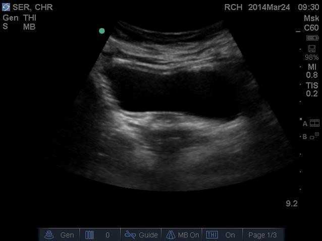
State Of The Art An Integrated Approach To Pelvic Floor Ultrasonography Santoro 2011 Ultrasound In Obstetrics Amp Gynecology Wiley Online Library
Computed Tomography Evaluation Of The Piriformis Muscle Axial Computed Download Scientific Diagram

Pelvis And Abdomen Radiology Key
Source : pinterest.com
